김일봉 (대구시 김일봉 내과 의원)

39세 남성 환자로 내원 5일 전부터 간헐적인 설사, 하복부 동통이 발생하였다.
내원 시 발열이나 오한은 없었다.
이학적 검사상 하복부 둔통이 있었다.
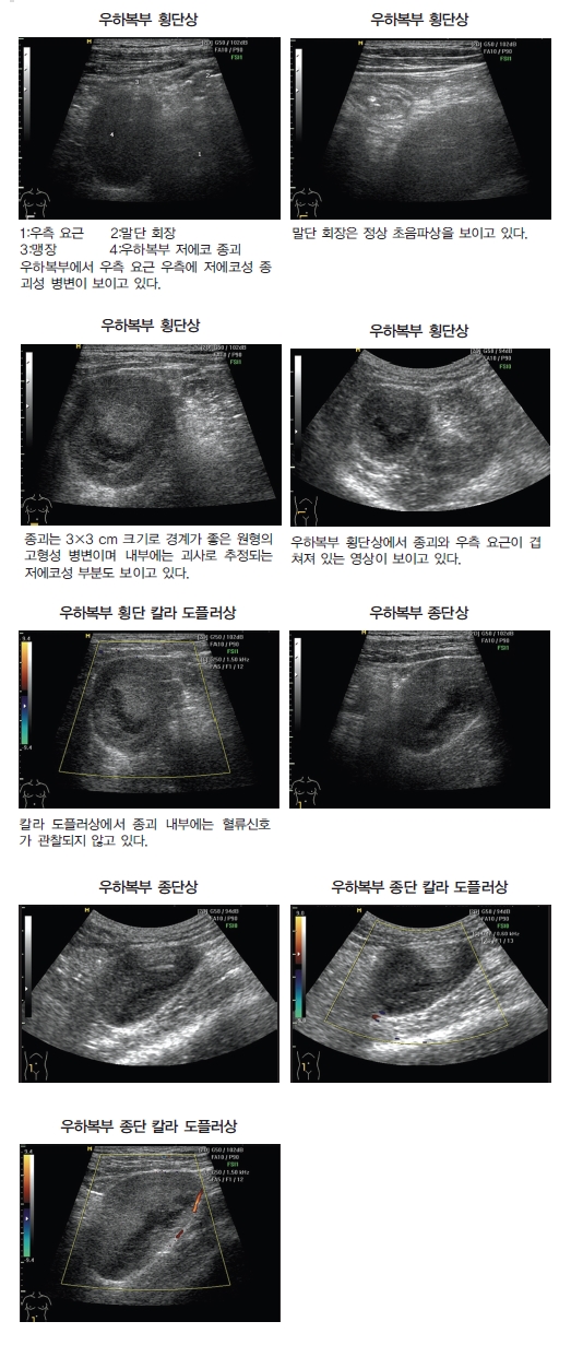
초음파 검사상 우하복 충수 주위 농양이 의심되어 파티마병원 일반외과로 전원하였다.
▶ CT of the abdomen and pelvis
Right iliopsoas muscle과 접하여 3×3.9 cm sized low density lesion 보인다.
Enhancement study상 거의 enhance 되지 않았다. Normal appendix 보인다.
Liver : small cysts at right lobe
GB and BILIARY TREE : multiple gallstones
PANCREAS : normal size and shape 보이며 focal lesion 보이지 않는다.
SPLEEN : mild splenomegaly
BOTH KIDNEY : normal size, shape 보이며 focal lesion 보이지 않는다.
ADRENAL GLAND : 특이소견 없다.
MESENTERY, OMENTUM : 특이소견 없다.
SMA, SMV : 특이소견 없다.
BOWEL LOOP : abnormal bowel wall thickening 보이지 않는다.
BLADDER : 특이소견 없다.
CONCLUSION : low attenuation lesion along the right psoas muscle.
R/O Primary retroperitoneal mucinous cystadenoma
R/O Lymphangioma
R/O Cystic mesothelioma
R/O Nonpancreatic pseudocys
▶ 대구시 파티마 병원 일반외과에서 개복수술을 시행하였다.
Gross : Submitted are 4 parts. The first one is a whitish soft tissue measuring 1×0.3 cm. The second one is a brownish lymph node measuring 0.6×0.3×0.3 cm. The third specimen is a gallbladder measuring 6×2×1.5 cm. On opening the lumen contains two irregilar shaped stones measuring 1 cm of the larger one. The mucosa is greenish velvety appearance without solid component. The fourth is a appendix measuring 7 cm in length and 0.6 cm in diameter. The serosal surface is unremarkable. On sectioning the body contains greenish fecaluth. Blocked in Aretroperitoneal mass, B-LN, C-gallbladder, D-appendix.
IMMUNOHISTOCHEMICAL STAIN :
S-100 : Positive (Positive in control block)
FINAL DIAGNOSIS :
Retroperitoneum, incisional biopsy : Neurofibroma
Lymph node, excisional biopsy : Reactive hyperplasia (0/1)
Gallbladder, cholecystectomy : Ch
김일봉 medifonews@medifonews.com
< 저작권자 © Medifonews , 무단전재 및 재배포금지 >
- 본 기사내용의 모든 저작권은 메디포뉴스에 있습니다.